
Private Testicular Ultrasound in Portsmouth & Southampton
Discreet assessment with same-day results
A detailed ultrasound assessment of the testes, epididymides and surrounding scrotal structures, helping investigate testicular pain, lumps, swelling and other scrotal concerns.
£199 · Same-day digital report · No GP referral required
Is Testicular Ultrasound Right for Me?
A private testicular ultrasound—also known as a scrotal ultrasound—provides detailed imaging of the testicles and surrounding scrotal structures.
You may wish to consider a testicular ultrasound if you have:
- A lump in or around a testicle
- Testicular or scrotal swelling
- Persistent testicular discomfort or aching
- A feeling of heaviness in the scrotum
- A change in the size or shape of a testicle
- One testicle that appears larger than before
- A suspected epididymal cyst
- A suspected hydrocele
- A suspected varicocele
- Previous inflammation or infection requiring reassessment
- A history of testicular injury
- Follow-up of a previously identified testicular or scrotal finding
- Concerns about the appearance or feel of your testicles
A testicular lump or persistent change should also be assessed by your GP, even if you arrange a private ultrasound.





What Does a Testicular Ultrasound Examine?
Your sonographer will perform a systematic ultrasound assessment of both testicles and the surrounding scrotal structures.
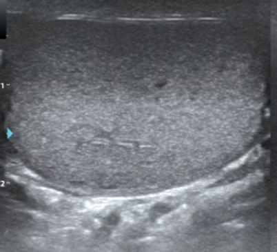
Testes
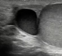
Epididymides
The epididymis is a small structure situated behind each testicle. Both epididymides are assessed for enlargement, inflammation, cysts or other visible abnormalities.
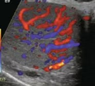
Testicular Blood Flow
Doppler ultrasound may be used to assess and compare blood flow within the testicles and surrounding structures.
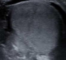
Scrotal Contents
The tissues and fluid surrounding the testicles are examined for visible abnormalities, including fluid collections such as a hydrocele.
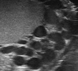
Varicocele Assessment
The veins within the scrotum may be assessed for enlargement consistent with a varicocele. Where appropriate, you may be asked to change position or gently bear down during this part of the examination.
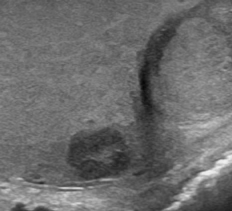
Palpable Lump or Area of Concern
If you can feel a specific lump or abnormal area, the sonographer will ask you to indicate its location so that it can be examined carefully and correlated with the ultrasound findings.
Why Patients Choose Us
Private diagnostics, run with NHS‑grade governance
- No GP referral required
- Findings explained during your scan
- Written report the same day
- Sonographer-led clinical governance
What to Expect
Before your scan, your sonographer will discuss your symptoms and medical history and ask you to identify any area of pain, swelling or a lump.
You will be given privacy to undress from the waist down and a covering to maintain your dignity. Ultrasound gel is applied to the scrotal skin and both testicles and surrounding structures are examined. Doppler ultrasound may also be used to assess blood flow.
The examination is usually comfortable, although tender areas may be sensitive. Your findings will be explained where appropriate, and your detailed digital report will be provided the same day.
Preparing for Your Scan
No special preparation is required. You can eat, drink and take your usual medication normally. Please wear comfortable clothing and be prepared to show the sonographer the exact location of any lump or area of concern.
Sudden severe testicular pain, particularly with nausea, vomiting or rapidly increasing swelling, requires immediate assessment in A&E and should not wait for a private ultrasound appointment.
What Happens if Something Is Found?
Many findings, such as epididymal cysts, hydroceles and varicoceles, are benign.
Any abnormality will be documented clearly in your report and explained where appropriate. Depending on the findings, you may be advised to share your report with your GP or urologist or arrange further assessment or follow-up.
Potentially significant testicular abnormalities require prompt medical or specialist assessment.
Limitations of Testicular Ultrasound
Ultrasound provides detailed assessment of the testicles and surrounding scrotal structures but cannot identify every cause of testicular or groin pain.
A normal scan does not exclude all causes of persistent symptoms, and ultrasound alone cannot provide a definitive tissue diagnosis or completely exclude testicular cancer.
If symptoms persist or worsen despite reassuring ultrasound findings, you should seek further medical assessment.